MRI Studies
Wake Radiology conducts one of the largest body MRI programs in the region. In addition to opening the first outpatient MRI center in Wake County, we were the first to perform many MRI procedures.
Wake Radiology opened the first outpatient MRI center in Wake County and the first to perform many MRI procedures. These include the first cardiac MR in Wake County as well as the first fetal MRI and contrast-enhanced 3D MRAs of the carotid, aorta, renal, mesenteric and peripheral arteries in Raleigh. Most recently, our radiologists were the first in the Triangle to perform an MRI procedure on a patient with an MRI-compatible pacemaker.
Fast Breast MRI
Abbreviated Breast MRI (also known as Fast Breast MRI) is a breast cancer screening tool available to women who have an average risk of breast cancer or who have dense breasts. Learn more about Fast Breast MRI at Wake Radiology.
MRI of the Chest, Abdomen and Pelvis
Some of the latest advancements in imaging have occurred in MRI of the chest, abdomen, and pelvis, or “Body MRI.” Tumors sometimes can be classified as benign or malignant solely upon the information provided by MRI.
Body MRI examinations are the most specialized of all MRI applications. All examinations require the patient to lie on his/her back for 30–70 minutes, as still as possible. Many body examinations require the patient to hold his/her breath, repeatedly, for up to 30 seconds.
If you are scheduled for a MRI of the chest, abdomen, or pelvis and have difficulty holding your breath, you should alert the MRI staff upon your arrival. Sometimes, coaching before the examination is successful in achieving an adequate breath-hold for imaging. However, some patients will find that they cannot hold their breath adequately, and an alternate imaging method, such as CAT (CT) scan or ultrasound, may be recommended instead of MRI.
In some cases, the radiologist may determine that the patient must be injected with contrast. This is not a sign that something is wrong, but merely that additional information is required/requested. An examination that requires contrast is not an unpleasant experience. All contrast agents are FDA-approved and have an extremely low incidence of allergic reaction.
Some patients may be injected with Glucagon, a synthetic hormone that reduces motion within the bowel for about 60 minutes. Bowel motion can cause significant blurring of images in the abdomen and pelvis, potentially rendering images useless. The injection is given in the muscles of the arm, much like an immunization. This drug may induce hypoglycemia (low blood sugar) in patients approximately 90 minutes after injection, causing some nausea, dizziness, and trembling. Because eating can prevent these symptoms nearly completely, patients are required to eat chocolate, or other forms of sugar, prior to leaving the facility. Patients are also instructed to eat a meal soon after the examination. Diabetics will typically not be given this injection, so please inform the staff if you have this condition.
MRI of the Brain and Spine
Almost all imaging of brain and spine involves the patient lying on his/her back for 30–60 minutes. In some cases, the radiologist may determine that the patient must be injected with contrast. This is not a sign that something is wrong, but merely that additional information is required/requested. An examination that requires contrast is not an unpleasant experience. The contrast material has an extremely low incidence of allergic reaction, and most patients do not even notice the injection.
Note: Due to recent internationally accepted evidence that Gadolinium injection is linked to the onset of a rare disease known as Nephrogenic Systemic Fibrosis (NSF), Wake Radiology adheres to the nationally mandated guidelines set forth by the FDA. We will not utilize Gadolinium for MRI studies on those patients who are at risk of NSF, such as those with documented severe chronic renal insufficiency or dialysis dependence. Those patients may safely have non-contrast MRIs if required clinically.
MRI of the Bones and Joints
Almost all imaging of bones or joints involves the patient lying on his/her back for 30–60 minutes. Occasionally, other positions are required to image small joints or special areas. In some cases, the radiologist may determine that the patient must be injected with contrast. This is not a sign that something is wrong, but merely that additional information is required/requested. An examination that requires contrast is not an unpleasant experience. The contrast material has an extremely low incidence of allergic reaction, and most patients do not even notice the injection.
Some special types of joint imaging require “arthrograms,” in which the patient is injected with contrast material inside the joint prior to MR imaging. Patients having these examinations will be instructed to arrive earlier than usual, so that a specially trained musculoskeletal radiologists may be present to administer the contrast into the joint.
MRI of the Blood Vessels (MRA, or MR Angiography)
When physicians need to see blood vessels, they create images called “angiograms.” Typical angiograms require admission to a hospital for the procedure, but MR angiograms can be performed without risk or hospitalization, in 30–70 minutes depending upon the body part to be imaged.
Patients undergoing MR angiography of any part of the body except the head will receive an injection of contrast material in the vein of an arm. In some cases, a second contrast injection may be required for some parts of the body. An examination that requires contrast is not an unpleasant experience. The contrast material has an extremely low incidence of allergic reaction, and most patients do not even notice the injection. MR angiography of the head (Circle of Willis, COW MRA) can be performed without a contrast injection.
Because very large amounts of data are created during these studies, they can easily have hundreds of images that require hours of manipulation to interpret. Referring physicians will be notified of results as soon as is possible.
How an MRI is Performed
MRI is an amazing technology that creates images for a radiologist to interpret from the water in your body. Giant magnets allow your body to receive radio waves and “echo” them back. A computer uses the information within the echoes that bounce back from your body to create images. The images created are unique to a patient, depicting his or her anatomy and any disease that may be present. The whole process is safe and painless. Some patients are so comfortable inside the magnet that they actually fall asleep while this advanced imaging takes place.
Anxiety-Free MRI Experience
At Wake Radiology, it’s easy to have a comfortable and anxiety-free experience during an MRI. Our MRI technologists have extensive experience in reducing patient stress, worry and anxiety during an MRI. We understand how important this imaging procedure is and we work closely with our patients to make them comfortable and help them complete the exam. To administer IV sedation your exam will be scheduled when our radiologists are available, and the patient will need to bring a responsible adult driver that must remain on-site for the duration of the exam.
After you arrive, at your request, we will place a small IV (intravenous) line into your vein, and through that we will administer a small amount of Valium. This will relax you for the exam, making it possible for you to easily manage your own stress or anxiety. We will not “put you to sleep,” but rather will relax you thoroughly for the duration of the examination.
Please note that if we give you Valium at our facility, you will need to have someone drive you home. Although you will feel nearly normal by the time the MRI is concluded, you will still be subtly affected by the medication, and cannot drive safely. Also, you should take the next few hours to relax, to allow the medication to wear off naturally.
MRI and Pacemakers
The Biotronik Pro MRI Eluna and Entovis Pacing Systems are the only pacemakers that can be scanned in our outpatient offices. Please verify the device and leads on your pacemaker identification card. Call our office if you have any questions, 919-232-4700 option 1.
MRI and Contrast Agent
In most cases an MRI exam does not require an injection. In some situations, however, a substance known as a contrast agent (Gadolinium) may be needed to enhance the ability of the MRI to see into your body. All contrast agents are FDA-approved and have an extremely low incidence of allergic reaction.
Note: Due to recent internationally accepted evidence that Gadolinium injection is linked to the onset of a rare disease known as Nephrogenic Systemic Fibrosis (NSF), Wake Radiology adheres to the nationally mandated guidelines set forth by the FDA. We will not utilize Gadolinium for MRI studies on those patients who are at risk of NSF, such as those with documented severe chronic renal insufficiency or dialysis dependence. Those patients may safely have non-contrast MRIs if required clinically.
The Safety of MRI
Magnetic resonance imaging is very safe. There are no health risks associated with the magnetic field or the radio waves used by the machine. Any metallic substance on your person can affect the quality of the diagnostic images. It can also cause discomfort or injury to you when placed in the magnetic field, and may exclude you from the exam. However, some special circumstances limit the use of a magnetic field, so it is important for you to tell us if any of the following apply to you or someone accompanying you into the exam room:
- cardiac pacemaker, defibrillator (AISCD) or artificial heart valve
- metal plate, pin or other metallic implant
- intrauterine device, such as Copper-7 IUD
- insulin pump or other infusion pump
- aneurysm clips
- previous gunshot wound
- middle/inner ear implant
- ever been a metal worker (had metal shavings or slivers in eyes)
- permanent (tattoo) eye-liner
- If you could be pregnant *
- artificial joints or metallic plates **
* Be sure to tell us if you are pregnant. Pregnant patients should discuss the examination with the radiologist prior to the appointment/examination. Although there are no known side effects on the developing baby, it is recommended that a pregnant woman wait until the second trimester for MR imaging. There are some exceptions to this rule. Wake Radiology also feels that Gadolinium (contrast material) should not be injected in the setting of pregnancy, and thus pregnant patients will only have non-contrast MRIs.
** Metallic Hardware Joints: You can safely undergo MRI if you have orthopedic metallic hardware in your joints—such as a metallic plate or hip replacement. However, if the metal device is located close to the part of the body being examined, the images can be seriously degraded and useless.
Our Safety of MRI Checklist:
Resources & MRI Checklist
Scheduling Your MRI
If you need an MRI, talk with your doctor about having your procedure at one of our MRI outpatient offices located in Cary, Chapel Hill, Fuquay-Varina, Garner, Holly Springs, Raleigh, Wakefield and Wake Forest. If you need to schedule an MRI, contact our MRI Scheduling Team at 919-232-4700, Patients option 2, MRI option 1.
MRI STUDIES
Anxiety Free MRI Experience
It’s easy to have a comfortable and anxiety-free experience during an MRI. Our MRI technologists have extensive experience in reducing patient stress, worry and anxiety during an MRI. We understand how important this imaging procedure is and we work closely with our patients to make them comfortable and help them complete the exam.
What to Expect During Your MRI
When a doctor orders an MRI, patients know they’re coming to find out more about their health concern or the extent of an injury. What they don’t know, is what to expect during the MRI exam. At Wake Radiology, your MRI experience is important to us. That’s why we’ve pulled together information about what to expect during and after your MRI exam.
Arriving at Your MRI Appointment
When you arrive, we ask you to check in at our front desk. We’ll confirm basic personal and health information and then give you an MRI safety checklist to complete. We are required to review this with every patient before entering the MRI scan room. It is important that we know about any implantable device or metal that is in or outside of your body, like aneurysm clips, pacemakers, cochlear implants, insulin pumps, neurostimulators or any injury to your eyes involving metal slivers or shavings.
One of our MRI technologists will walk you to a changing room where you will change into scrub pants and a scrub top or a gown. It’s important that patients remove all clothing prior to their MRI exam. We ask patients to remove:
- All outer clothing, including shoes
- Bras or any undergarment that could have metal in it. Most underwear may be worn.
- Glasses
- Jewelry – watch, earrings, necklace, bracelets, etc.
- Hairpins, barrettes or other hair accessories containing metal
- Purses, wallets, cell phones
All personal items are placed in a locker and secured. Before walking to the exam room, we’ll review your medical history and your MRI safety checklist again, confirming that you are not wearing anything that contains metal.
Our Safety Checklist Process
Our Safety Checklist Procedures
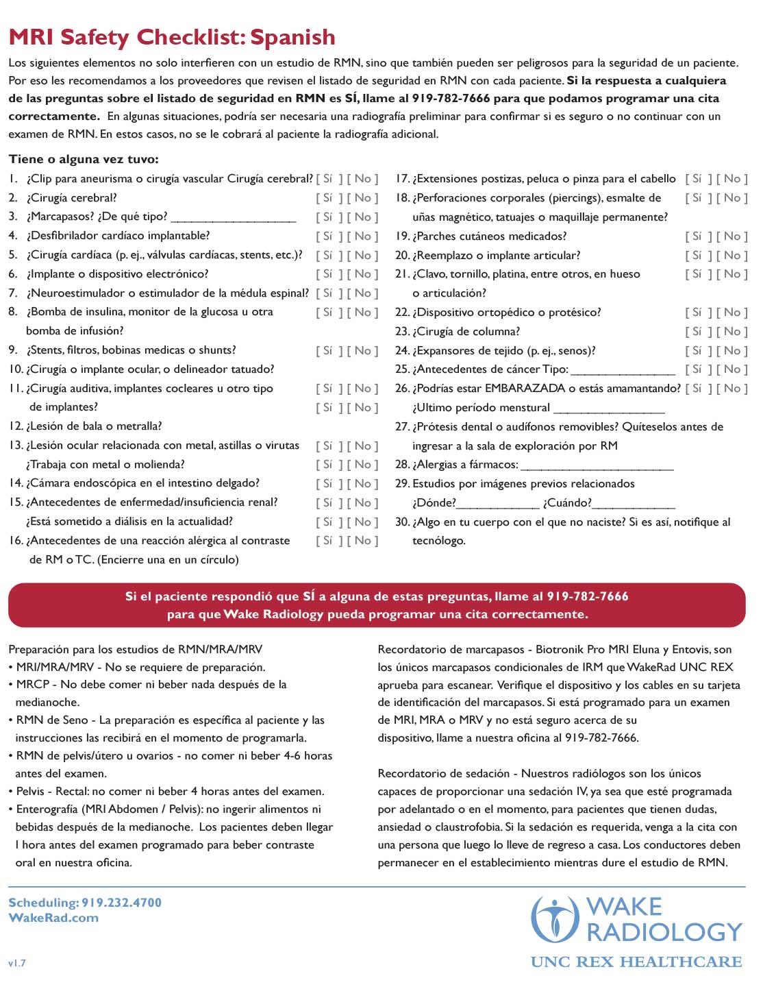
Wake Radiology understands the power of the MRI magnet and takes extra steps to ensure that patients are safe to enter the scanner. Prior to the appointment, Wake Radiology will review the MRI safety checklist with each patient. In some situations, a preliminary X-ray may be necessary to confirm whether or not it is safe to continue with an MRI exam.
| MRI Safety Checklist (English) | MRI Safety Checklist (Spanish) |
 |
 |
As we enter the exam room, we’ll confirm the procedure that your doctor ordered and fully explain what is about to happen. Some exams require patients to go into the MRI machine head first, while others enter feet first. It’s important that we get the body part that we’re going to image into the center of the magnet. This ensures we obtain the best possible images of that section of the body.
You will lie down on a moveable table during your procedure and you may have a blanket, if desired. An imaging device called a coil may be placed around the area of the body that will be imaged. The coil is a plastic framework that cradles the head, joint or body during the exam and helps us obtain better quality images. Depending on the type of exam that is ordered, you may need an MRI contrast material to further detect or diagnose potential abnormalities. If so, the technologist will place an IV in your arm to administer the MRI contrast.
Next, we will give you a pair of headphones and an emergency call bell to hold. These items ensure you can hear the MRI technologist during the entire exam and communicate if there is any concern. We will explain how the exam is made up of small scans during which you will need to hold as still as possible and answer any final questions you may have. We’ve found that most patients, even those who are extremely nervous, are more comfortable when they know exactly what to expect. Our technologist will remain in the room until you are properly positioned and comfortable.
At Wake Radiology, it’s easy to have a comfortable and anxiety-free experience during an MRI. Our MRI technologists have extensive experience in reducing patient stress, worry and anxiety during an MRI. We understand how important this imaging procedure is and we work closely with our patients to make them comfortable and help them complete the exam. To administer IV sedation your exam will be scheduled when our radiologists are available, and the patient will need to bring a responsible adult driver that must remain on-site for the duration of the exam.
When you are ready for the exam, the technologist will walk into an adjacent room that has a large window that looks into the MRI room. Our technologists can see the patient during the entire MRI exam. The technologist then confirms that you can hear them and will continue to update and coach you throughout the procedure. We routinely tell patients exactly what is happening and how quickly they are moving through the exam. Time between scans can vary as the technologist reviews the images and sets up the next scan. This is normal and no reason for alarm.
During the MRI, you will hear knocking or buzzing sounds from the machine. Again, this is normal. These knocking noises only last while images are being taken. Your headphones or earplugs help dampen these sounds. Your MRI technologist will communicate with you after each scan. Once the exam is complete, the technologist will come back into the exam room to help you out of the machine and escort you to the changing area. You will change back into your own clothes and are now free to go.
Getting Your Results
Once the MRI is complete, your images are immediately routed to one of our subspecialty trained radiologists for interpretation. For example, if you had a knee MRI, we make sure that your images are interpreted by one of our musculoskeletal/orthopedic radiologists. If previous images are available, we obtain these and our doctor compares the current exam with this historical information. This helps us determine changes and how those should factor into a diagnosis. Our radiologist then provides an actionable report to your doctor who ordered the MRI. Your doctor will follow up with you regarding your results and develop a treatment plan based on that diagnosis.